"I have used a wide variety of secondaries and Jackson ImmunoResearch has consistently been the best. The fluorophores are bright and stable and their selective (x reactivity removed) secondaries have always shown species specificity in multiple labeling."
Janet Duerr, Ohio UniversityRating: 5.0
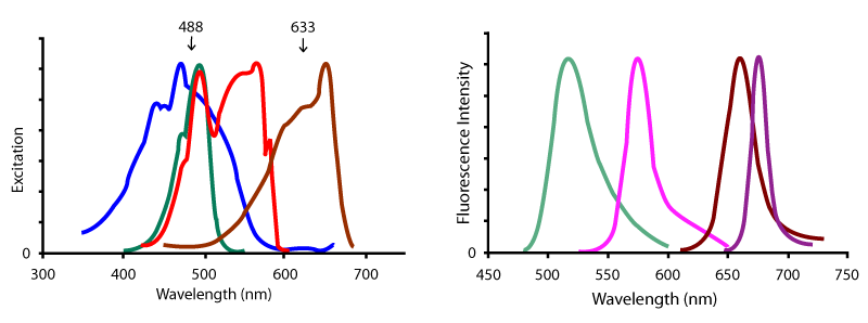
Peridinin-Chlorophyll-Protein (PerCP) is a 35.5 kDa fluorescent peridinin-chlorophyll-protein complex isolated from dinoflagellates. Jackson ImmunoResearch offers the form found in Dinophyceae sp. It has a broad spectrum of excitation with the main peak at 482 nm, and a long Stokes shift to an emission peak at 677 nm. PerCP conjugates are large complexes suitable for cell surface labeling techniques such as flow cytometry.
PerCP is available conjugated to:
| Excitation Peak (nm) | Emission Peak (nm) | |
|---|---|---|
| Peridinin-Chlorophyll-Protein (PerCP) | many, 488 | 675 |
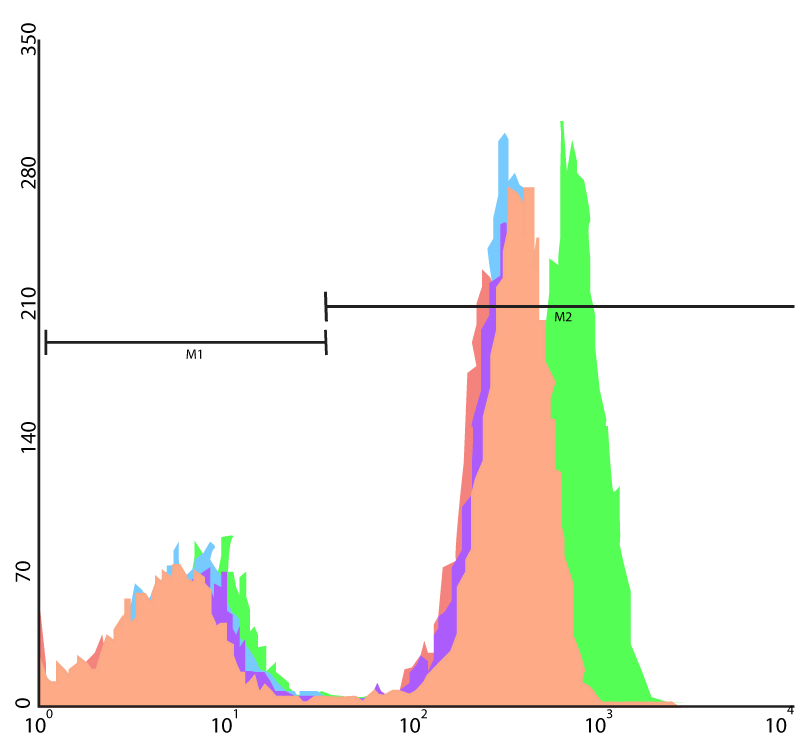
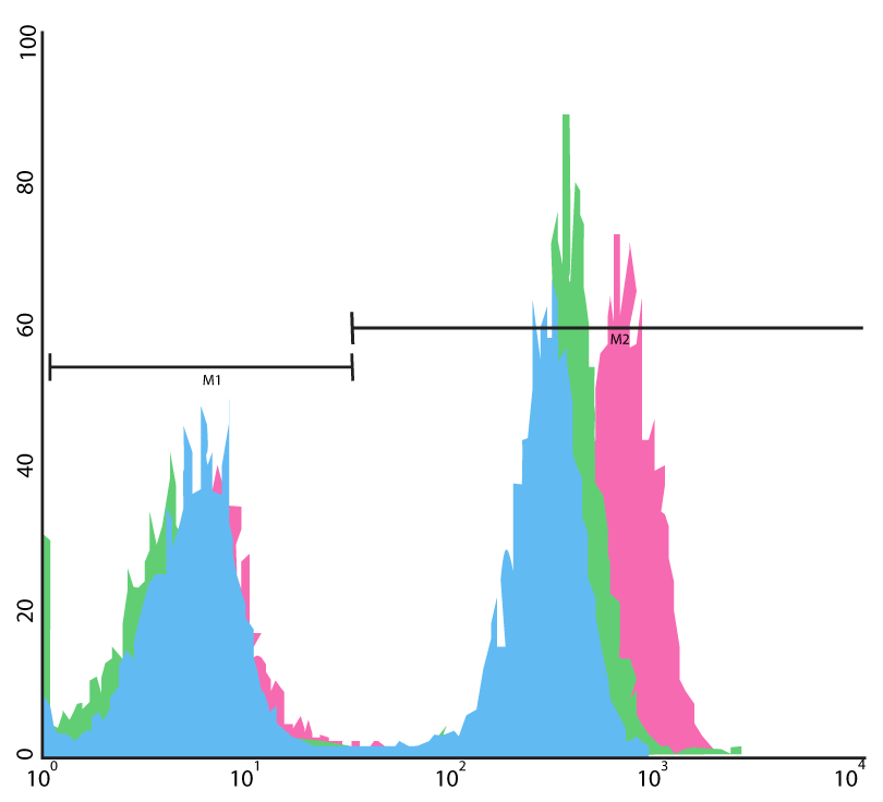
PerCP conjugates of highly adsorbed secondary antibodies are offered to label unconjugated primary antibodies, and PerCP-streptavidin is offered to label biotinylated primary or secondary antibodies (Figures 2 and 3). Two practical labeling protocols are possible with the products.
Compared with a single-step PerCP-conjugated primary antibody (Figure 1), about the same level of fluorescence is obtained with a two-step procedure using a biotinylated primary antibody and PerCP-conjugated streptavidin. A consistent, slightly higher signal is achieved by using an unconjugated primary antibody and PerCP-conjugated secondary antibody. Although three-step procedures are usually undesirable for flow cytometry, a somewhat greater amplification may be obtained with unconjugated primary antibody, biotinylated secondary antibody, and PerCP-conjugated streptavidin (Figure 2).
PerCP conjugates may be used alone or with Alexa Fluor® 488 (or FITC) and R-PE for one- to three-color analyses with a single-laser flow cytometer equipped with an argon laser emiting at 488 nm. Up to four-color analyses with low compensation are easily achieved by adding APC-conjugated antibodies with 633 or 635 nm excitation provided by a dual-laser flow cytometer (Figure 3).


Use the spectra viewer below to compare PerCP with other fluorophores, along with instrument specifics (laser/filter) for suitability in your assay.